Request a callback
Contact us
Get Direction
Zehra Manzil, G-3, 91, Lady Jamshedji Rd, near Paradise cinema, Mahim West, Mumbai, Maharashtra 400016
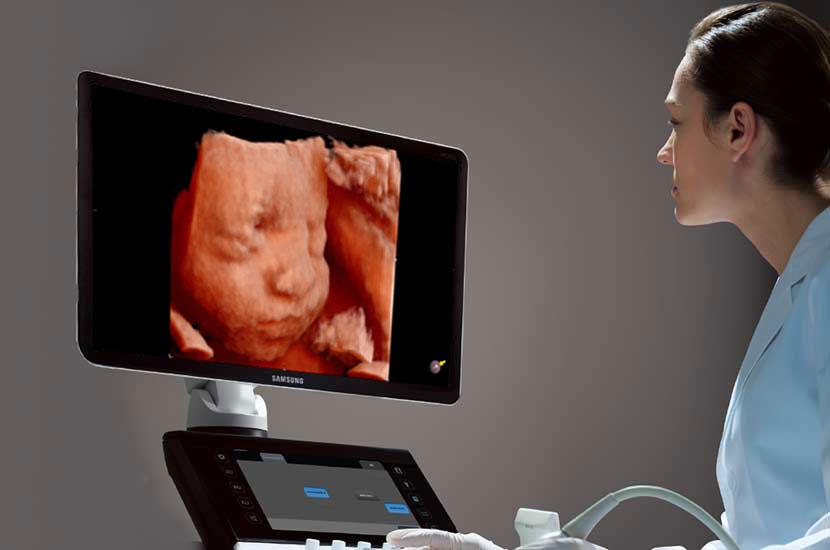
What is 3D Ultrasound/4D Ultrasound?
A three-dimensional i.e. 3D ultrasound is one of the most advanced techniques of ultrasound in which a standard, two-dimensional greyscale ultrasound image is converted into a volumetric, three-dimensional image.
With the help of post-processing techniques and AI (artificial intelligence), the acquired image is enhanced and can be viewed in all the three-axis for a detailed examination. This technique has been developed in order to solve issues related to obstetrics and gynaecology thereby reducing the dependence on ultrasonography test and also avoid operator related insufficiencies.
A four-dimensional i.e. 4D ultrasound is a real-time, dynamic ultrasound that complements 2D and 3D examination. By use of 4D ultrasound, the medical practitioner can assess dynamic images of the foetal face, respiratory movements, swallowing, movements of the lips and mouth, blinking of eyes and limbs movements.
A 3D ultrasound can be useful in gynaecology for the detection of uterine anomalies such as Mullerian duct abnormalities, to check the position of the intrauterine contraceptive device (IUCD), to locate and characterize uterine fibroid, endometrial polyps. It may also be useful in imaging adnexal lesions such as cysts, exophytic fibroids etc.
Preparation for 3D Ultrasound/4D Ultrasound
- Nothing in particular is required for routine 3D sonography or 4D sonography scans.
- Patients are advised to remove jewellery, belts etc which may obscure the part to be examined.
- Patients are always advised to bring previous reports for comparison.